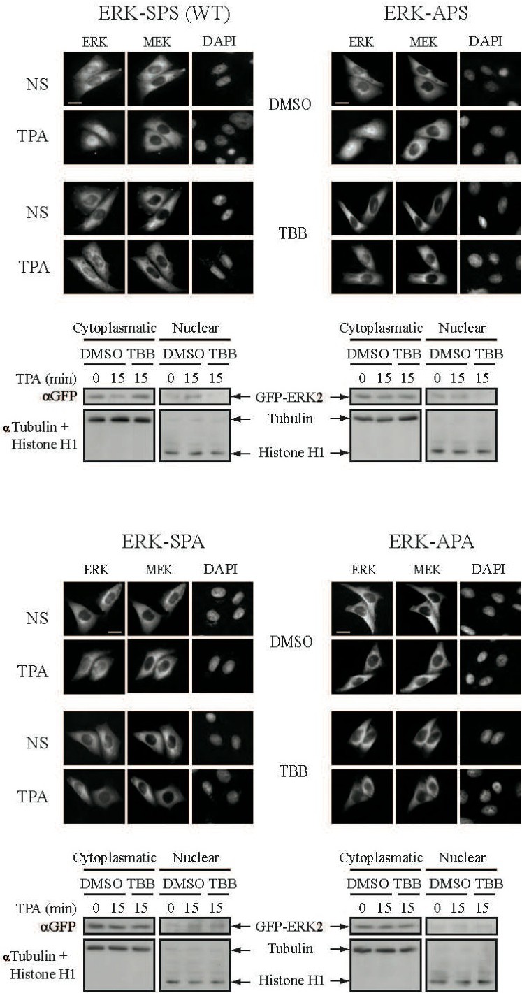
Fig. 6. Role of distinct Ser phosphorylation within the NTS in the nuclear translocation of ERK. CHO cells were co-transfected with the following GFP-ERK2 constructs: SPS (WT), APS, SPA, APA and RFP-MEK1. Thirty-six h after transfection the cells were serum starved, pretreated with either 10μM of TBB (10 μM) or DMSO control for 2 h, and then either stimulated with TPA (250 nM; 15 min) or left untreated, NS). The cells were processed and stained as in Fig.1A. Scale bar - 15nm. In the parallel experiment the cells were seperated to cytoplasmatic/nuclear fractions, and the amount of GFP-ERK2 constructs was analyzed with αGFP Ab (upper panels). Tubulin and Histone H1 served as loading control for cytosolic/nuclear fraction respectively (bottom panels).